May 14, 2020
K-State researcher develops cell-based MRI contrast agent for efficient cancer diagnosis

A cell-based magnetic resonance imaging, or MRI, contrast agent can efficiently light up the tumor area, providing crucial diagnostic information for cancer therapy, according to a Kansas State University study recently published in two Royal Society of Chemistry journals, Biomaterial Science and the Journal of Material Chemistry B.
"Our findings suggest that an engineered cell-based MRI contrast agent can provide clinically effective contrast at the site of the tumor or vasculature at a minimal dose thereby differentiating surrounding tissues," said Santosh Aryal, assistant professor of chemistry and principal investigator of the study. "This is a safer and much needed alternative to existing MRI contrast agent."
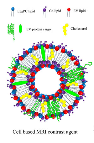
According to the study's lead author, Sagar Rayamajhi, a graduate student in chemistry, cells release small vesicles, called extracellular vesicles, or EVs, to communicate with each other. The researchers have isolated these vesicles from the immune cell and reconstructed them with contrast agents for efficient cancer diagnosis.
"Due to immune cell origin, these vesicles acquired the properties of immune cell that naturally seeks for disease or inflammatory sites, therefore, reconstructed vesicles can stay for a longer time in blood vasculature to target cancer or disease sites," Rayamajhi said. "We found that by reconstructing the MRI contrast agent in the vesicle system, the contrast enhancement ability increased by more than double. This will allow us to reduce the dosage of clinically used contrast agents about two to three-fold while maintaining contrast in the clinical window."
Rayamajhi said this discovery is important because the current agent used in MRIs is based on gadolinium metal, which can pose a safety issue. The researcher's proposed cell-based MRI contrast agent may be a good alternative for diagnostic imaging.
"The beauty of this study is the optimized use of surface functionalization strategies to engineer magnetic functionalization in EVs while maintaining its membrane integrity," Aryal said.
There are various methods — physical, biological or chemical — to change the surface of extracellular vesicles to add a desired function. Each method comes with specific advantage and disadvantages.
"There is no perfect method," Aryal said. "But, if we choose smartly a combination of two approaches, then, maybe we can get what we want. And, that’s what we did here by combining chemical strategy to synthesize a lipid probe with magnetic contrast agent gadolinium and physical strategy to incorporate the lipid probe in the lipid bilayer of EVs. This is a beautiful combinatorial approach that incorporated magnetic property in EVs while retaining its structural and functional integrity."
In addition to Aryal and Rayamajhi, co-authors include graduate student Ramesh Marasini, chemistry graduate alumnus Tuyen Nguyen, Brandon Plattner, pathologist and clinical associate professor at the Veterinary Diagnostic Lab and David Biller, radiologist and professor of clinical sciences in College of Veterinary Medicine.
This research was supported by the Johnson Cancer Research Center and University Small Research Grants and made possible with the new clinical MRI 3Tesla system in the College of Veterinary Medicine. The researchers add a special thanks to Scott VanAmburg, MRI and radiologic technologist, Veterinary Health Center for his assistance in MRI experiments.
The researcher's next goal is to use a similar cell-based MRI contrast agent for the early mapping of metastasis to improve the cancer survival rate.
