Veterinary researcher validates human tracheal/bronchial-cell model for influenza A infections of trachea
Friday, March 13, 2015
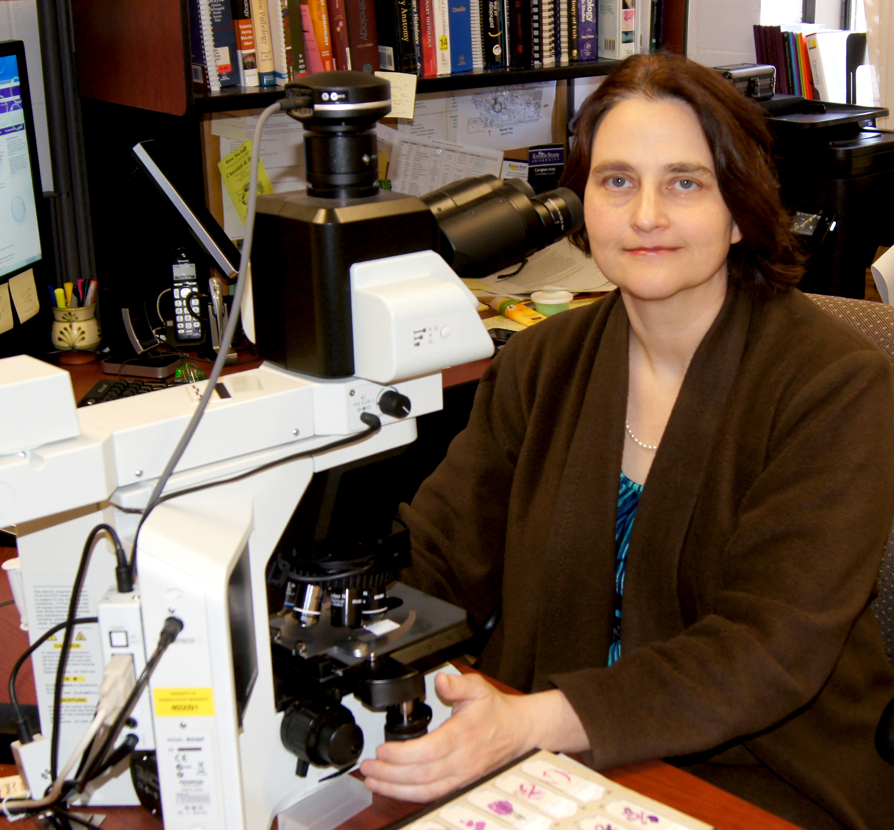
MANHATTAN — A recently hired veterinary pathologist is bringing multiple skills to her new position in the College of Veterinary Medicine at Kansas State University, including experience with a human primary cell line used for modeling human influenza infections.
A. Sally Davis is an assistant professor of experimental pathology in the diagnostic medicine and pathobiology department. Part of her appointment at Kansas State University is to provide pathology support to the Department of Homeland Security's Center of Excellence for Emerging Zoonotic Animal Diseases at the university.
Davis graduated in 2014 from the National Institutes of Health Comparative Biomedical Scientist Training Program in partnership with North Carolina State University. At the NIH, she conducted her doctoral research under Dr. Jeffery K. Taubenberger in the Laboratory of Infectious Diseases at the National Institute of Allergy and Infectious Diseases. Her work was funded through the Intramural Research Program of the National Institute of Allergy and Infectious Diseases. Her research has produced a paper on an in vitro model for influenza infections that will be published in May's Journal of Histochemistry & Cytochemistry.
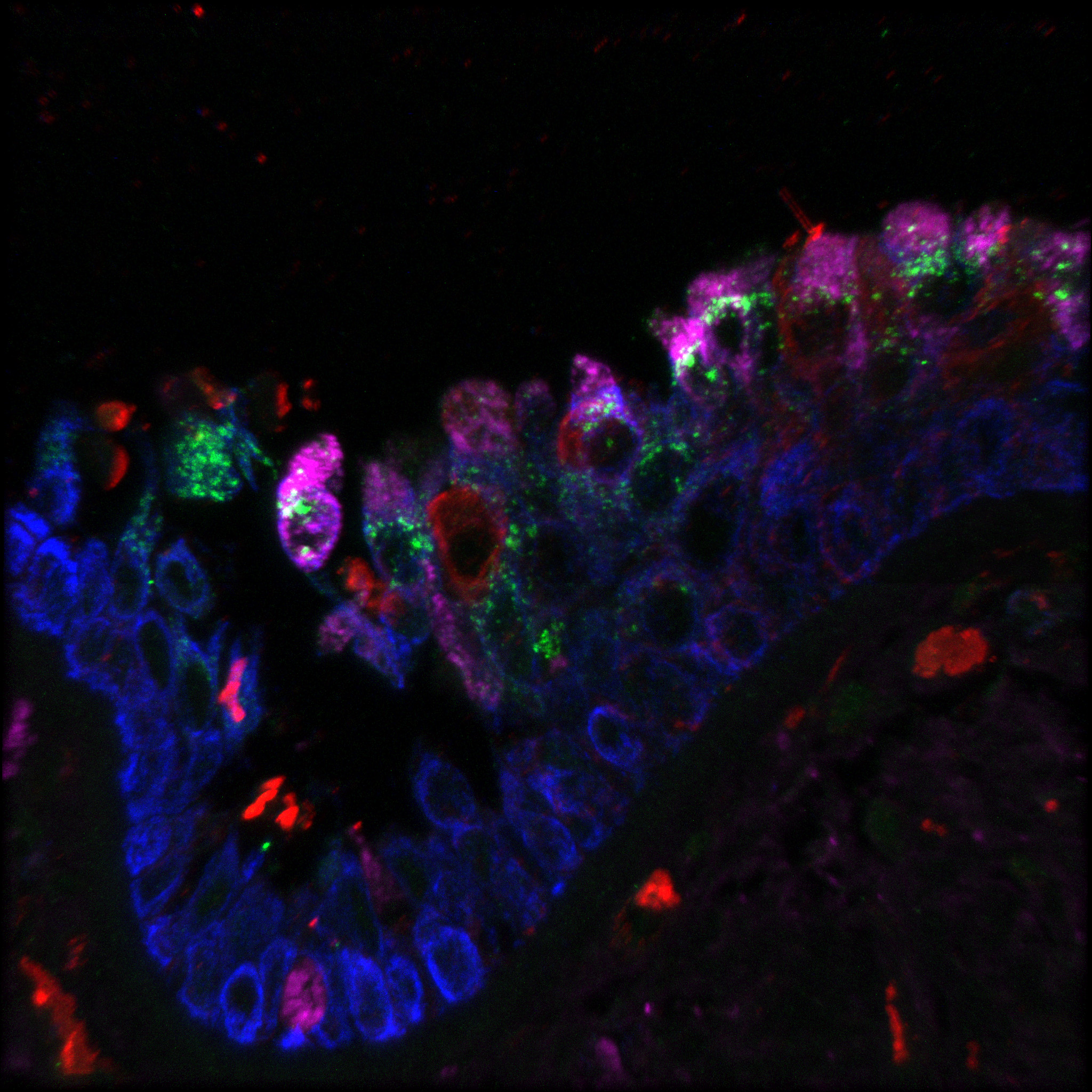
"This is a human cell line that has been in use for quite a while in respiratory research," Davis said. "However, there was no detailed comparison of the epithelium that these cells can form in the laboratory to their source tissue, the human trachea. Therefore, the idea was to perform morphological comparisons as well as study differences in response to influenza infection of the 'pseudo' trachea versus human trachea sampled from autopsy material, including individuals who died from an influenza infection. We found that the model was particularly strong at early infection time points, but has limitations at later time points."
The results will enable respiratory virus researchers to better interpret data generated with this system. Additionally, this type of research provides an approach to validation of other in vitro models derived from other animal species.
"It is important to know both the strengths and limitations of these models so that scientific findings may be accurately interpreted," Davis said.
Part of the research used electron microscopy and immunofluorescence confocal microscopy, which are areas of expertise Davis will use at Kansas State University. She said she will present guest lectures and assist in collaborative research projects using the microscopy facilities.
Davis also will present a paper on the 1918 pandemic influenza — Spanish flu — at the Experimental Biology conference in Boston at the end of March. For this work, Davis has won the Histochemical Society-sponsored American Society of Investigative Pathology travel award and has been invited to co-chair the "Microbial Interactions with the Host" mini symposium at the conference.
In her research, Davis said she enjoys working with and mentoring students. At the Kansas State University she anticipates working with both undergraduate and Doctor of Veterinary Medicine students, including publishing with students on future research papers.
"I was originally a high school biology teacher, so I've always had a strong interest in working with students," Davis said. "Even while I was working on my own Doctor of Veterinary Medicine at North Carolina State University, I formally tutored some of my classmates. During, my residency training, I mentored DVM students in pathology and research projects."
While much of her recently published work has involved primarily human applications, Davis chose to return to a veterinary medicine environment so that she could better focus on the animal-human interface of infectious diseases. She looks forward to working on nonhuman projects, such as vaccine efficacy studies in swine and sheep, and building new collaborations at the university while she continues her previous collaborations with the NIH.


