Kansas State University researcher receives one of 30 new NSF EPSCoR fellowships
Wednesday, Sept. 27, 2017
 and Jocelyn McDonald")
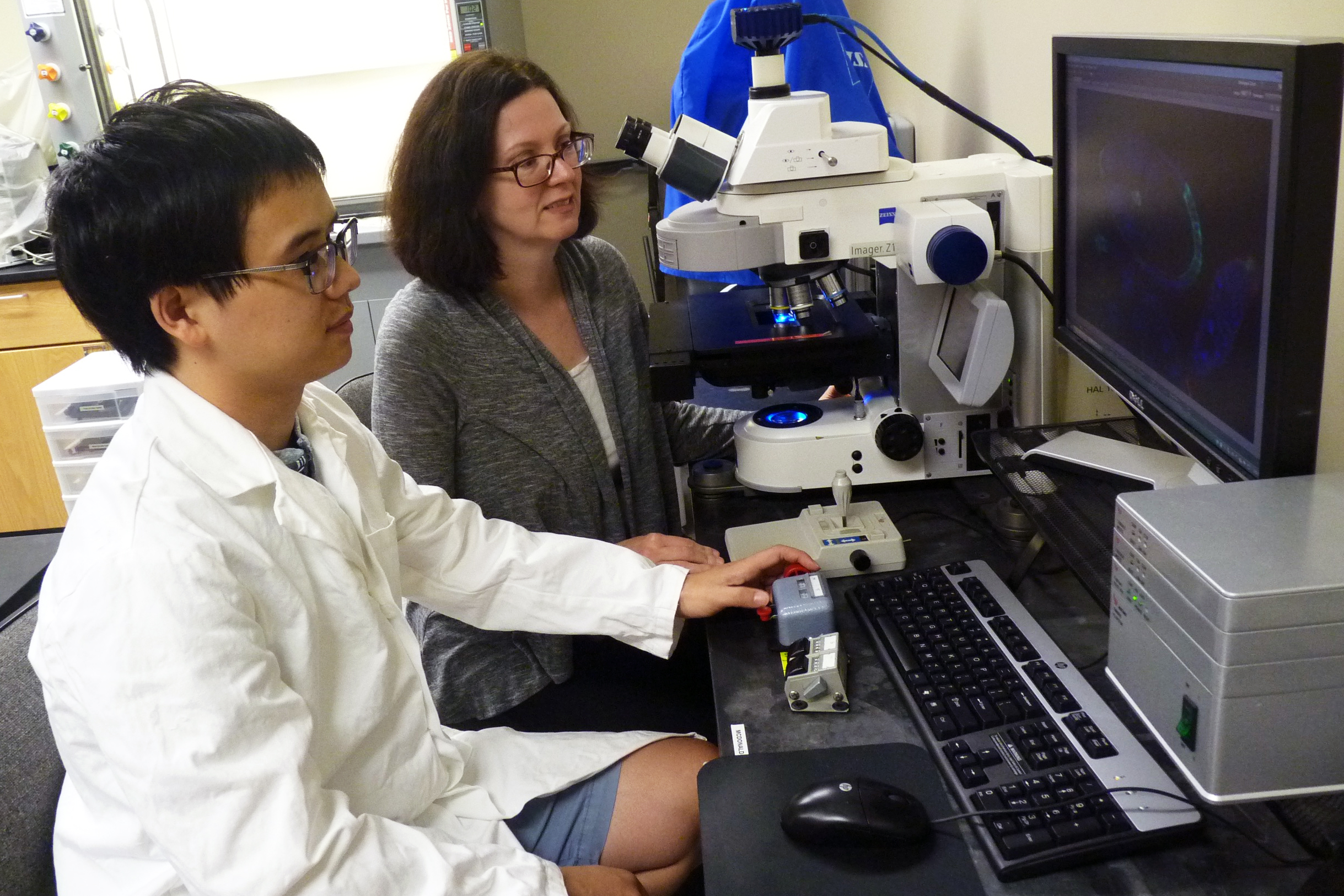
Division of BiologyAssistant Professor Jocelyn McDonald, right, who is one of 30 awardees of a new National Science Foundation fellowship, and Yujun "Eugene" Chen, left, postdoctoral researcher, work with fluorescent images of embryonic cells. | Download this photo.
MANHATTAN — A Kansas State University researcher has been awarded an inaugural National Science Foundation fellowship to learn new microscopic methods to take 3-D movies of live cells as they move.
One of 30 researchers nationwide, Jocelyn McDonald, assistant professor in the Division of Biology, was awarded the new Research Infrastructure Improvement Track-4 fellowship through the National Science Foundation's Established Program to Stimulate Competitive Research, or EPSCoR, initiative.
The fellowship provides opportunities for pre-tenure investigators to further develop their individual research potential by partnering with another researcher at a premier research center. McDonald will collaborate with Denise Montell at the University of California, Santa Barbara to learn how to use light sheet fluorescent microscopy, which can capture 3-D images of live cells as they move in tissues.
"Often, we think of cells as being still and not doing much, but many cells move from one place to another in a tissue or an organ," McDonald said. "This is really important in making fully formed embryos, healing wounds and immune system functioning."
McDonald works with specific fruit fly cells that migrate as a group, or collective, which is broadly important for the embryonic development of many organs. According to McDonald, it is not well understood how these cells stay ordered and migrate inside dense tissue. Light sheet fluorescent microscopy could help scientists see that process in more detail and for a longer time than previous techniques.
"There are many techniques to look at cells in their native environment, but usually the light from the microscope is too intense," McDonald said. "This particular system, the light sheet microscope, has gentle light, it's three-dimensional and we can do really long-term imaging to view the cell group on its four- to six-hour journey in the tissue and make 3-D movies."
Another method that McDonald and an accompanying postdoctoral researcher, Yujun "Eugene" Chen, will learn as part of the fellowship is a technique using a confocal microscope's strong light that can turn proteins on and off in the cells to control cell movement.
"We already have some genetic evidence that certain proteins are very important in the migration of these cells," McDonald said. "We don't know if they are required during the whole journey or only parts of the journey. By turning on a specific protein at different parts of the journey, I hope we can answer that question."
The two-year fellowship for nearly $177,000 includes funds for travel, salary support for McDonald and Chen and some supplies. At the end of the two years, McDonald and Chen will be expected to share what they have learned with others at Kansas State University.
"This technique could be used in a variety of biological disciplines, not just development biology," McDonald said. "It can do live cell imaging — without killing the cell — of plants, insects or vertebrates."
