
Updated: 02 January 1999
E-mail: Steve J. Upton
Division of Biology, Kansas State University, Manhattan, KS 66506
go to the Cyclospora
homepage or
Biology
546 tutorial
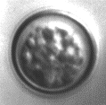
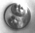
Unsporulated oocyst in early stages of sporulation during contraction of
sporont (cytoplasm). The globules throughout the cytoplasm will gradually
increase in number, fuse, and split into the refractile bodies
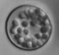
Unsporulated oocyst in early stages of sporulation during contraction of
sporont (cytoplasm). The globules have become more numerous throughout the
cytoplasm will soon coalesce to form one large globule
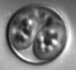
Unsporulated oocyst in early stages of sporulation during contraction of
sporont (cytoplasm). The globules in the cytoplasm have coalesced to
form one large globule
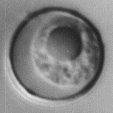
Unsporulated oocyst. The large globule is now beginning to split into the
refractile bodies
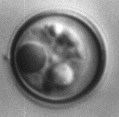
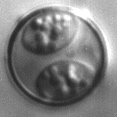
Partially sporulated oocyst. The sporocyst walls have formed, but the
sporozoites have not developed
Fully sporulated oocyst with sporocysts. A small amount of oocyst
residual material remains
Fully sporulated oocyst compressed slightly due to coverslip pressure

Unsporulated oocyst (slightly lower magnification than photos above)
demonstrating autofluorescence at 340-380 nm (adapted
from the CDC
parasite image library)

Home | Search | What's
New | Help | Comments
Kansas State University | Biology Division