Teacher Tips

This experiment can be used to investigate the following concepts and phenomena:

1. To learn how to grow yeast, to change their environment, and to observe them through the microscope.

2. To observe and learn to identify the characteristic shapes (morphology) of yeast cells at important stages of their sexual life cycle.

3. To follow the inheritance of a color trait (pink colony vs. cream colored colony) and see if one variation is dominant and the other recessive, or whether intermediate colors are inherited.

The yeast life cycle may be used to teach a variety of concepts. (See the Cycles in the Life of Yeast and A Simple Cross and Cell Division Cycle segments in video tape I.) As the students gain skills in manipulating the yeast you may wish to have them go through the cycle several times using different haploid strains. This particular protocol uses HA2 (pink) and HBT (cream) haploid strains. Microscopes are used to observe the characteristic shapes of the stages of the yeast life cycle. Color changes are used to develop an understanding of dominant and recessive phenotypes.

You may wish to show a variation on genetic behaviour by crossing various strains. For example, HA2 (pink) and HB1 (pink) produce cream colored diploids when the two haploids are mated. It is possible to use these strains to demonstrate that when gametes combine, the offspring sometimes look different than either parent.

Whan HA2 & HB1 mate, they mark the transition between haploid and diploid with a color change (haploids pink; diploids cream). This feature may be useful if microscopes are not available to examine the cell shape change that also marks the transition from haploid to diploid.

Getting Ready:

You may wish to prepare this first set of plates for the students. Yeast strains usually come from the supplier growing on agar slants. Contamination may be a problem when students use the master set of yeast strain slants as the source of their strains. A quick method for preparing the "Getting Ready" plates is for the teacher to make a master plate, incubate it overnight, and then use the replica plating method to make copies for the students. (See Replica Plating segment in video tape III.)
Student Data Record Sheet
If you want your students to do the "Getting Ready" step of the experiment, you can subculture the strains on to YED plates. One subculture plate of each mating type will supply enough yeast for all the students. If several groups need access to the yeast at the same time, you may want to make several subculture plates (See video tape segment Subculturing Yeast.)

You may wish to show your students the video tape segment Subculturing Yeast which demonstrates the use of sterile toothpicks for moving yeast cells.
If you collect the used toothpicks in small beakers, the toothpicks can be rinsed, brushed, and resterilized in an autoclave or pressure cooker and used again.

Click here to return

Teacher Tips

Mating Two Haploid Strains and Observing Zygotes:

1. This mating procedure is illustrated in video tape segment Monohybrid Cross.

4. If your students keep a lab journal you may wish to have them record data and drawings as journal entries. An alternative method is to copy and hand out the Data Record Sheet provided with these materials.

5. In order to view the cell shapes, the students need to use the high power lens of their microscopes; 400X is adequate. If streaming of the cells in the water currents under the coverslip is a problem you may wish to seal the coverslip with fingernail polish. (See video tape segment Wet Mount Slides.) You can display the slides on a video monitor using a video microscope. It is possible to use a home video camera and TV monitor for this purpose. (See Microscope and Home Video Camera segment in video tape III.)

You can use a microscope to observe cells directly on the agar surface by gently dropping a cover slip onto the area to be observed. Focus the microscope through the cover slip just as you would on the slide. Naturally, the plate is almost certain to become contaminated!

Click here to return

Teacher Tips

Time Shifting:

Rather than refrigerating all the plates after 3 hours, you may wish to prepare a refrigerated mating mixture in advance for the students to use to make their mating mixture wet-mount slides.

If you have a "premated" mixture available, the students should be able to complete "Mating Two Haploid Strains and Observing Zygotes" in one 50 minute lab period. If your students are expected to observe their own mating mixtures, you will need to refrigerate their plates so they will be able to observe the characteristic mating shapes the next day. You will also need to adjust the time line for your class.

The time line assumes that you have prepared a premated mixture for the students to observe on the same day that they make their own mating mixture. The point to remember as you work out the logistics for your class is that it takes about three hours at 30o C for mating to occur and for zygotes to become visible. Refrigeration will delay the process.

Click here to return

Teacher Tips

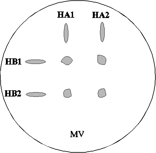
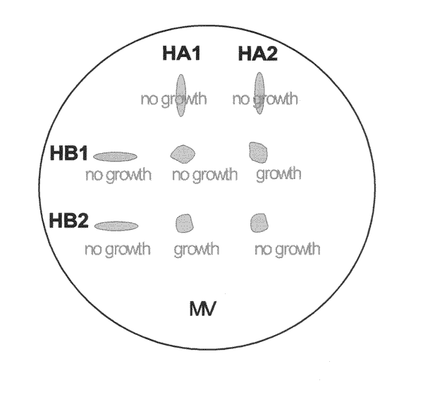
Selecting the Diploids:

In this experiment the students use complementation to select diploid cells. HA2 carries the ade2 mutation but it carries a functional TRP5 gene. It is not able to synthesize adenine due to the ade2 mutation but it is able to synthesize tryptophan. The absence of adenine in the growth medium prevents HA2's growth on MV. HBT carries a functional ADE2 gene but it carries the trp5 mutation. It is not able to synthesize tryptophan but it is able to synthesize adenine. The absence of tryptophan in the growth medium prevents HBT's growth on MV. When the two haploid cells mate and fuse, the resulting diploid cell has one functional copy of the ADE2 gene and one functional copy of the TRP5 gene. It is able to synthesize both adenine and tryptophan thus allowing the diploid to grow on MV medium.

Genotype of haploid parents

mutant functional
HA2 ade2 TRP5
HBT trp5 ADE2
Genotype of diploid cells
Each diploid cell has one functional copy of each gene.

HA2/HBT TRP5/trp5 ade2/ADE2

Selecting the diploids:

You may wish to use the replica plating technique for this step. (See Replica plating segment in video tape III)

Click here to return

Teacher Tips

Numbers refer to steps in the student procedure.

Presporulation:

1. The diploid cells growing on MV should be cream-colored.

Sporulating the Diploids:

1. The diploid cells growing on YED should remain cream-colored.

2. Since the diploid cells don't divide rapidly on YEKAC, be sure to transfer enough cells so that you have plenty to produce asci. For some diploid strains, sporulation efficiency is affected by cell density. If you don't find asci in one section of the streak, check sections of the streak with higher and lower cell densities.

Click here to return

Teacher Tips

Numbers refer to steps in the student procedure.

Observation of Asci and Germination of Spores:

2. Some strains take longer than three days to sporulate. The diploid cells produced in the HA2 x HBT cross tend to sporulate quickly. For example, if you put the diploid cells on YEKAC on Friday you should have some spores by Monday.

4. This technique is routinely used to spread cells out far enough so that a single cell can produce a single isolated colony. The ascus wall is tough and tends to hold the spores together. In many cases even after spreading the cells, there will still be more than one spore stuck together. As noted in the experimental procedure, some of the spores will be mating type a and some mating type . After they germinate, they may mate and produce diploid colonies. If both the spores happen to carry the ade2 mutation, the resulting diploid will be homozygous for ade2 and have a pink phenotype. In other cases, the spores may be too far apart from one another to mate. These spores will form haploid colonies. If the spore carries the ADE2 allele of the gene, the colony will be cream-colored. If the spore carries the ade2 allele of the gene, the colony will be pink. You may wish to pick samples of the pink colonies, subculture them overnight on YED and then transfer them to YEKAC as a test of ploidy. Diploid cells will sporulate and haploid cells will not.

Looking for the Missing Color:

The reappearance of the pink phenotype demonstrates that the complete sexual life cycle has been completed.

Click here to return

Teacher Tips

Objectives and Applications:

Our understanding of the nature of genes at the molecular level came from the concept of allelism, that mutations can occur at many places in a gene and there are multiple genes that contribute to the same phenotypic trait. Mutation to a red colony color have been found in two different genes in the biosynthetic pathway for adenosine monophosphate (AMP), ade1 and ade2. When students can understand these relationships, the can appreciate the concept of allelism and how we learned what genes are. (See The Two Gene Hypothesis and A Genetic Test For Allelism segments in video tape I).

Objectives:
1. To make the four possible crosses between two different red mutants and determine the color and growth requirements of each of the diploids.
2. To determine whether the results support the hypothesis that the two red mutants have mutations in different genes.
3. To see that this experiment provides a test for whether two mutations affect the same or different genes (a test for allelism).

Subculture Parent Strains:
You may wish to prepare this first set of plates for the students. Yeast strains usually come from the supplier growing on agar slants. Contamination may be a problem when students use the master set of yeast strain slants as the source of their strains. A quick method for preparing these subculture plates is for the teacher to make a master plate, incubate it overnight, and then use the replica plating method to make copies for the students. (see Replica Plating segment in video tape III.) These student copies need to be incubated overnight before the strains are mated.

If you want the students to do this step of the experiment, you may wish to subculture the strains on YED plates. One subculture plate of each strain will supply enough yeast for all the students. If several groups need access to the yeast at the same time, you may want to make several subculture plates of each strain. (See Subculturing Yeast segment in video tape III.)

Click here to return

Teacher Tips

Numbers refer to steps in the student procedure.
    Cross all four strains:
  1. Remind the students to use a new sterile toothpick for each different strain and to keep the spots from touching each other. If individual students don't get the expected results from the crosses keep in mind the possibility that they may have cross contaminated the strains during this step.
  2. If the haploid cells don't get thoroughly mixed there will be areas of unmated cells on the edge of the mating mixture. These unmated areas will show the phenotype of the haploid parent rather than the phenotype of the diploid cells. For thorough mixing the mating mixture should cover an area larger than the two spots of haploid yeast.

Click here to return

Teacher Tips

The students are asked to record only color and growth on MV. They will not be confirming the mating phenotype of the diploid cells. It is possible to take the diploid cells produced in this exercise through the life cycle if you want to follow the segregation patterns of mating types, ADE1 and ADE2.

The expected data supports the two gene hypothesis. A functional copy of both genes is necessary to produce the normal cream colored colonies. The genes code for different enzymes in the same biochemical pathway (AMP synthesis).

Figure 4 and Figure 5

Click here to return

Teacher Tips

Complementation, allelism, and defining a gene:

Genetics is the study of genes. Genes carry the information that defines every organism. To accomplish this, genes (with the help of the cells they live in) must do three things:

1. They must reproduce: their information must be copied faithfully.
2. They must be transmitted: their information must be passed precisely to new cells.

3. They must act: their information must result in metabolic reactions and cellular structures.

Let's think about a life cycle experiment where you cross two red haploid strains together and get a diploid that is cream colored. When you sporulate the diploid you recover both red and cream colonies. A model (theory) developed by Gregor Mendel to explain inheritance of different traits in peas might explain these results. It is called the recessive-dominant theory. Suppose that in every normal yeast cell there are two genes--for now call them GENE1 and GENE2--that are needed for the colonies to be cream colored. If one of the red strains had a mutant form of one of the genes and the other strain had a mutant form of the other gene, we could explain the results of the cross and make some predictions that you could test in another experiment. Let's assume that the mutant genes are damaged forms of the normal ones and for now call the mutant forms gene1 and gene2. We could say that one of the red parent strains contained GENE1 and gene2 and the other parent gene1 and GENE2. The diploid formed between them could then be represented as follows:

GENE1 gene2
gene1 GENE2

From this you can see that there is one copy of each of the normal genes (capital letters) and one copy of each of the mutant genes (lower case letters). If the normal genes still work in the presence of the mutant genes, they can still do whatever it is that makes the colony cream colored. The mutant genes are just going along for the ride.

Let's see if there is some way we can test this model. What would happen if you crossed two red haploid strains that had the same mutant gene, such as crossing a gene1 GENE2 with another gene1 GENE2. Then the diploid would be:

gene1 GENE2
gene1 GENE2

This diploid has no copies of the normal GENE1, so we would expect it to be red. In the same way, if you crossed GENE1 gene2 by another GENE1 gene2 strain you would get

GENE1 gene2
GENE1 gene2

which has no copies of the normal GENE2, so it should also be red. To summarize this, we can say that when mutations affect different genes, whethere the same phenotype or not, they will complement each other; when crossed together the diploid will have the normal phenotype. This demonstrates that they are not alleles. When they are in the same gene, they will fail to complement each other and that is taken as evidence that they are alleles. So complementation provides a genetic test for allelism and gives a criterion for determining whether two mutations affect the same or different genes, and therefor, different functions. Complementation also provides a tool for selecting diploids from a mating mixture. This tool was used in A Simple Cross.

Comments on genetic nomenclature:

The following tables show the shorthand strain numbers, the genotypes in the official Yeast Genetics nomenclature, and a description of the phenotypes for the strains used in this experiment. This experiment, which shows that mutations in two different genes can give similar phenotypes, illustrates why this more elaborate nomenclature is necessary.

HAPLOID STRAIN NO. GENOTYPE PHENOTYPE
HA1 MATa ade1 ADE2 pink, will mate with MAT
HA2 MATa ADE1 ade2 pink, will mate with MAT
HB1 MAT ade1 ADE2 pink, will mate with MATa
HB2 MAT ADE1 ade2 pink, will mate with MATa

DIPLOID GENOTYPE PHENOTYPE
HA1 /HB1 MATa/MAT ade1/ade1 ADE2/ADE2 pink, will not mate with either mating type, will not grow on MV
HA1/HB2 MATa/MAT ade1/ADE1 ADE2/ade2 cream colored, will not mate with either mating type, will grow on MV
HA2/ HB1 MATa/MAT ADE1/ade1 ade2/ADE2 cream colored, will not mate with either mating type, will grow on MV
HA2/HB2 MATa/MAT ADE1/ ADE1 ade2/ade2 pink, will not mate with either mating type, will not grow on MV

click here to return

Teacher Tips

Objectives and Applications:

This experiment is designed to be used in conjunction with the traditional textbook treatment of a simple dominant/recessive two-factor cross. It gives graphical realism to the ever-popular Punnett's Square. (See A Traditional Dihyrid Cross segment in video tape I).

Objectives:
1. To see first-hand how the genotypes of the haploid gametes combine to form the phenotypes of the diploid offspring for dominant and recessive alleles.

2. To see that there is no uncertainty in the outcome (phenotypes of the offspring) when there is no uncertainty in the genotypes of the gametes. In the diploid dihybrid cross the uncertainty is the result of random sampling of the pool of gametes from the parental cross.

Yeast strains usually come from the supplier growing on agar slants. Contamination may be a problem when students use the master set of yeast strain slants as the source of their strains. You may wish to subculture the strains on YED plates. One subculture plate containing all eight haploid strains will supply enough yeast for a class. If several groups need access to the yeast at the same time, you may want to make several subculture plates (See Subculturing Yeast segment in video tape III). Alternatively you may wish to prepare the Day 1 plates for the students. A quick method for preparing these plates is for the teacher to make a master plate, incubate it overnight, and then use the replica plating method to make copies for the students (see Replica Plating segment in video tape III).

See the Laboratory Methods section for agar media recipes. You will need to make the plates several days in advance.

Each student or team will require at least 50 sterile toothpicks for this experiment. Toothpicks are sterile from the box and there are 750 toothpicks in each box. (See Laboratory Methods F: Using Toothpicks and Inoculating Loops)

Each student or team will require at least 1 mating grid. A copy master of six grids is included with these materials.

1st Day:
Have the students write the expected genotypes and phenotypes on the mating grid and then tape the grid to the bottom of the YED plate as a pattern for plating the yeast.
Only the eight haploid strains are plated on Day 1.

2nd Day:
Stress that fresh toothpicks should be used for each different strain or mating mixture. The best results are obtained when only small amounts of yeast are used to make each mating mixture.

3rd Day:
Once again the best results are obtained when only small amounts of yeast are transferred during replica plating. If you use replica plating equipment you will need a fresh sterile velvet for each plate that is copied.

4th Day:
If the mixtures were not mixed thoroughly there may be pink areas in otherwise cream-colored mixtures. These mixtures should be scored as cream-colored.

Comments on genetic notation: We have chosen to use traditional notation for the genotypes in this experiment so that it will be more consistent with usual textbook treatments of the dihybrid cross and Punnett's Square. In later experiments we will introduce the more sophisticated notation of modern yeast genetics. Even so, you have some options for how much you want to simplify things.

Case 1

We can represent the hypothetical dihybrid cross between two diploids as follows:

RRtt x rrTT

But in yeast, we don't actually cross diploids. We first sporulate these diploids, obtaining two types of spores, each in both mating types:

ARt, ArT and BRt, BrT

These represent the gametes in the parental cross. We then mate either ARt x BrT or BRt x ArT to obtain the F1 diploids, which would be:

F1: ABRrTt

If we sporulate this diploid, we get the following eight haploid genotypes (spores or gametes) shown on the previous page.

In this experiment the students start here to obtain the F2 generation, without the complication of statistical sampling, by setting up the following grid of crosses:

F2:
ART ARt ArT Art
BRT c+ c+ c+ c+
BRt c+ c- c+ c-
BrT c+ c+ r+ r+
Brt c+ c- r+ r-

where c = cream, r = red, + = grows on MVA, and - = doesn't grow on MVA Case 2 In yeast, mating type is controlled by the mating type genes that segregate as alternative Mendelian alleles, so it seems natural to include them in the formal description of the genotypes, as we have done above. However, it may simplify the discussion to treat mating type separately. The A's and B's could, therefore, be omitted in all the above genotypes. If, for example, you describe a cross as Rt x rT, it is implicit that they are different mating types. If this approach is taken, then the whole scheme simplifies to the following:

P1: RRtt x rrTT

gametes: Rt and rT

F1/P2: RrTt

F2:

RT Rt rT rt
RT c+ c+ c+ c+
Rt c+ c- c+ c-
rT c+ c+ r+ r+
rt c+ c- r+ r-

Comments:
The main difference between this experiment and the analogous case in higher organisms, is that in yeast we have control of the life cycle so that we can deal directly with the haploid stages. We can isolate, as pure strains, the haploid parents of the F2 generation, which correspond to the gametes from the P2 parents. By being able to identify these, isolate them as pure strains, and make all the possible crosses, we remove all the chance factors. In higher organisms, we hypothesize the segregation of alleles in a hybrid when it goes through meiosis. If we make two assumptions, 1) that the alternative alleles segregate independently into the gametes, and 2) that there is dominant/recessive expression of the respective alleles, then we can predict the 9:3:3:1 ratios among the F2 offspring. Accordingly, this experiment provides a direct demonstration of how the diploid phenotypes are derived from the genotypes of the parents.

This does not demonstrate independent segregation, which requires showing that the haploid phenotypes did, in fact, occur in equal numbers among the spores from the F1 diploids.

In the context of animal breeding, or even human genetics, what we do with yeast when we isolate two haploids and mate them has a parallel in in vitro fertilization. However, we do not yet have the ability to score many genetic traits in gametes of higher forms, nor can we propagate them asexually.

Click here to return

Teacher Tips

Genetic test for Allelism

Objectives and Applications:


This is a variation on Traditional Dihybrid Cross and Two Genes/One Trait experiments. It can serve as a briefer substitute for both or it can be used as a supplement. In the former case you may want to introduce some of the ideas developed in those experiments. (See The Two Gene Hypothesis and A Genetic Test For Allelism segments in video tape I).

Objectives:
1. To have the students see how the pattern of phenotypes is altered in a dihybrid cross when both mutations yeild the same phenotype compared with the traditional case of two different phenotypes.

2. To see how complementation provides a way to distinguish between non allelic mutants that have the same phenotype.

Yeast strains usually come from the supplier growing on agar slants. Contamination may be a problem when students use the master set of yeast strain slants as the source of their strains. You may wish to subculture the strains on YED plates. One subculture plate containing all eight haploid strains will supply enough yeast for a class. If several groups need access to the yeast at the same time, you may want to make several subculture plates (see Subculturing Yeast segment in video tape III). Also, you may wish to prepare the 1st Day plates for the students. For a quicker preparation of these plates, the teacher can make a master plate, incubate it overnight, and then use the replica plating method to make copies for the students (see Replica Plating segment in video tape III).

See the Laboratory Methods section for agar media recipes. You will need to make the plates several days in advance.

Each student or team will require at least 50 sterile toothpicks for this experiment. Toothpicks are sterile from the box.

Each student or team will require at least 1 mating grid. A copy master of six grids is included with these materials.

Day 1:
Have the students write the expected genotypes and phenotypes on the mating grid and then tape the grid to the bottom of the YED plate as a pattern for plating the yeast.

Only the eight haploid strains are plated on Day 1.

Day 2:
Stress that fresh toothpicks should be used for each different strain or mating mixture. The best results are obtained when only small amounts of yeast are used to make each mating mixture.

LDay 3:
Once again the best results are obtained when only small amounts of yeast are transferred during replica plating. If you use replica plating equipment, you will need a fresh sterile velvet for each plate that is copied.

Day 4:
If the mixtures are not mixed thoroughly there may be pink areas in otherwise cream-colored mixtures. These mixtures should be scored as cream-colored.

The pattern that results should represent the Punnett Square as it is used to diagram the dihybrid cross. The pattern of dominance of ADE1 and ADE2 over ade1 and ade2, respectively, should also be clear. This case is slightly different from the usual textbook dihybrid , since the two mutant genes involved, ade1 and ade2, have the same phenotype (red, adenine-dependent). Note how different the patterns of phenotypes is form the two different phenotype case, in spite of the same underlying segregation pattern. You can also use this as an example of the red phenotype at one locus being epistatic to the cream phenotype at the other locus.

When red, adenine-requiring mutants were first discovered and studied, it was this test that demonstrated that there are two genes involved. This allelism test is the basis for assigning and newly discovered mutant to an already known gene.

In this experiment the students start with F1 haploids (gametes) to obtain the F2 generation, with no statistical sampling complications by setting up the following grid of crosses:

F2:

A++ A1+ A+2 A12
B++ c+ c+ c+ c+
B1+ c+ r- c+ r-
B+2 c+ c+ r- r-
B12 c+ r- r- r-

where c = cream, r = red, + = grows on MV, and - = doesn't grow on MV

Click here to return

Teacher Tips

Environmental effects on Colony Color

Objectives and Applications:
This experiment can be used to investigate the following concepts and phenomena:

Numbers refer to steps in the student procedure.
Materials: 1. If HA2 or HB2 is not available, you can substitute any of the following adenine mutant strains: HA1, HB1, HA12, HB12, HAR, HBR. The yeast should be subcultured overnight on a YED plate. (See Subculturing Yeast segment in video tape III.)

2. YED agar plates may be substituted for MV agar plates in this experiment. However, since YED agar provides a small amount of adenine, the pink lawn will grow all the way to the edge of the plate without a section of no growth. It may also take longer for the pink color to develop.

The procedure for this experiment is illustrated in the Effect of Adenine on Red Mutant Yeast segment in video tape II.

1. Any sterile container will work for mixing the yeast cell suspension such as baby food or other types of jars. It is possible to recycle them for this purpose by washing and sterilizing them in an autoclave or a pressure cooker. You can also purchase presterilized plastic specimen containers from a science supply vendor.
The amount of water in relation to the yeast cells is not critical as long as the suspension is visibly turbid. It takes about 1 milliliter of suspension for each plate, so multiply 1mL times the desired number of plates. The best results are obtained by using a fresh yeast suspension, but yeast cells are relatively stable in water so you might store the suspension in the refrigerator and use it for several days if necessary.

4. The surface water must be absorbed by the agar before the adenine disk is placed on the surface. If there is surface moisture, the adenine will diffuse in an uneven pattern across the surface instead of moving uniformly through the agar. The best results are obtained when freshly poured plates are allowed to sit at room temperature for 3 or more days before use.

Click here to return

Teacher Tips

Numbers refer to steps in the student procedure

5. Sterile forceps may be used to handle the adenine disk. The forceps may be sterilized by wiping them with an alcohol wipe instead of using the traditional flame sterilization method.

6. Note that the plates are incubated with the agar side down in order to keep the adenine disk in place. Usually plates are incubated with the agar side up in order to prevent any condensation from forming on the lid and dripping onto the yeast colonies.

Observe and Respond:

Development of growth and color rings:

1. How can you explain the white growth near the center where the adenine concentration is excessive? The answer to this question will vary based on the background of the students and the context in which you are using this experiment. What seems to be happening here is that the adenine concentration is high enough to support growth and also high enough to turn off the adenine synthesis pathway by feedback inhibition. Red pigment is only produced when the adenine synthesis pathway is turned on but it is blocked by an ade1 or ade2 mutation.
Do these white cells require adenine? Yes, this is an environmental difference, not a genetic change.
How can you test your answer experimentally? Move some of the white cells to YED agar without an adenine disk. Cells that do not require adenine will grow as cream colored colonies on YED and cells that still carry the red mutation will grow as pink colonies on YED. (See the next section "Retest the white yeast".)

2. Describe the nutritional conditions in the red zone. There is enough adenine to support growth but not enough to shut off the adenine synthesis pathway.

3. Why is there no growth at the edges of the plate? The adenine concentration is too low.

Click here to return

Teacher Tips

Retesting the white yeast:

1. Streaking for single cells is illustrated in the experimental procedure. This method is useful for examining the range of cell types in a sample. An alternative method, the single streak subculture method, will be adequate for this experiment if you wish to use fewer plates and have several students test their yeast on the same YED plate. (See the Streaking For Single Cells and Subculturing Yeast segments in video tape III)

Analyze your observations:

1. Explain whether the change from red to white near the center of the plate was a genetic or an environmental effect? The white yeast from the center of the adenine disk plate should grow as pink colonies on YED. The change from pink to white is environmental.

2. Give an explanation for this change or suggest another experiment you might do to learn more about it. In the YED environment, there is enough adenine to support growth but not enough to shut off the adenine synthesis pathway, so the cells form pink colonies. In the MV+ adenine environment, there is enough adenine to support growth. The concentration is also high enough to shut off the adenine synthesis pathway, so the cells form cream-colored colonies. One possible experiment would be to plate the white yeast from the first MV+ adenine plate onto an MV plate. If the red to white change was genetic, the yeast should now be able to grow on a plate with just MV agar. If the change was environmental, the white yeast should not be able to grow on just MV.

Click here to return

Teacher Tips


Effect of anaerobic growth on pigment formation:

1. If HA2 or HB2 is not available, you could substitute any of the following adenine mutant strains: HA1, HB1, HA12, HB12, HAR, HBR. The yeast should be subcultured overnight on YED agar. (See Subculturing Yeast segment in video tape III)

Growing the red strain anaerobically:

The procedure for this experiment is illustrated in the Anaerobic Growth of Red Mutant Yeast segment in video tape II.

1. See the previous section Effect of Adenine of Red Mutant Yeast for a discussion of making suspensions of yeast cells.

Click here to return

Teacher Tips

Numbers refer to steps in the student procedure

4. Most of the surface water should be absorbed by the agar before the agar is cut and folded. The best results are obtained when 1 mL or less of yeast cell suspension is placed on each plate and the freshly poured plates are allowed to set out at room temperature for 3 or more days before use.

7. Note that in order to keep the agar "sandwich" together, the plate is incubated with the agar side down. Normally, plates are incubated with the agar side up in order to reduce condensation problems.

Click here to return

Teacher Tips

Numbers refer to steps in the student procedure

Exposing the cells to air(O2):

1. The cells outside the "sandwich" should be pink and the cells inside the layers should be cream-colored. It is likely that bubbles of carbon dioxide gas will form between agar layers. 5. After about one hour the cream colored cells that have been exposed to the air will start to develop a pink color. The cells remaining between the agar layers should still be cream-colored. The responses to the following questions will vary according to the background information that has been given to the students. The following responses are examples.

Analyze your observations:

1. Is there any part in the sandwich where the yeast has not turned pink or red? Cream-colored inside, pink outside. 2. Is there a reason to think that the yeast growing there might be anaerobic? It is common for colored compounds to be formed by oxidation. If oxygen is not present (anaerobic conditions), the color does not develop. The lack of pink pigment in the cells between the agar layers supports the idea that anaerobic conditions were present between the agar layers.

3. Did you see any gas bubbles between the layers of agar? Bubbles of gas are formed between the agar layers. If so, can you think of an explanation for them? The yeast cells produce CO2 gas as a waste product during their normal growth. This helps keep the environment between the agar layers anaerobic.

4. What conclusion, if any, can you draw from this experiment about the role of oxygen in the formation of the red pigment? Oxygen is necessary for the formation of the red pigment.

5. Explain whether the change from white to red was a genetic or environmental effect? The effect was probably environmental. The yeast that stayed between the agar layers remained cream-colored, but still formed the pigment when grown aerobically. If the effect was genetic, the change would not be reversed for all cells.

6. Give an explanation for this change or suggest another experiment you might do to learn more about it. This was an environmental effect produced by the pigments formed in the cells by a reaction involving the oxygen in the air. The yeast that turned pink could be replated between agar layers to see if the change was genetic. If it turned once again cream-colored between the layers and then turned pink when exposed to air, the idea of an environmental effect would be supported.

Click here to return
Return to contents

Teacher Tips

Yeast Mating Pheromone

Objectives and Applications:


This remarkably simple experiment gives students a view of cellular physiology that is difficult to find. It also provides them with an introduction to a power research tool -- the bioassay -- that they can use to explore a wide range of phenomena. (See The Yeast Pheromone, -Factor segment in video tape I).

Materials:
To reduce contamination it is usually best to limit student use of the master set of yeast strains. You may wish to make one YED subculture plate for each mating-type on the previous day before the students start work. Students can then use those plates when they do their work on 1st and 2nd days. One plate of each mating-type will provide enough yeast for a whole class.

2nd Day:
A suspension containing cells at approximately 1 x 10e7 cells/ml will appear very cloudy.

Before you do this lab, you should test your microscopes' ability to view yeast cells directly on the plate. You will most likely need to remove the Petri dish lid and get the objective lens very close to the agar surface. At a short working distance, some lenses fog up and obstruct the view.

To overcome this problem you can drop a coverslip onto the area you wish to observe. Naturally this will most likely contaminate the plate.

It is possible to buy synthetic alpha factor. (-Mating Factor, Sigma Chemical Co.)
If your microscopes can't view cells directly on the agar plates, you may wish to add the synthetic alpha factor to mating-type a cells which are growing on agar or in liquid culture, and then use those cells to make wet mount slides for viewing.

Click here to return

Teacher Tips

Yeast Transformation

Objectives and applications:

The natural color trait of the ade1 mutant makes possible this simple, economical genetic transformation procedure. Students will see how the phenotype is a consequence of the genotype from a new, molecular prospective. The transformed cells produced will also make possible the follow-up experiment on plasmid loss, which illustrates the role of the centromere. (See Yeast Transformation segment in video tape I).

Materials notes:
- Plasmid DNA/carrier mixture: prepared as described in Schiestl and Gietz, 1989; this is enough for one transformation.
- MV medium: 1 plate for each experimental condition.
- Sterile pipettes: types for student use will depend on what materials you premeasure for the students.
- Spreaders: The use of alcohol to flame sterilize spreaders is dangerous and requires close supervision. Presterilized paper clip spreaders are much safer.
- Strain HA1L (ade1): one subculture plate will supply enough yeast for 4-5 students or teams. Plates can be grown several days in advance and stored in the refrigerator. Or, streak to isolate single colonies with sterile toothpicks and incubate 3-4 days until colonies are well grown. Use 3-4 colonies to make the yeast/lithium acetate suspension.

- Sterile Solutions: prepare working solutions from stock solutions just prior to use. Use distilled water and autoclave stock solutions and working solutions.

    Working solutions:

    Stock solutions:

Click here to return

Transforming the cells:

Procedure:
2. Be as accurate as possible given your pipetting equipment. One drop is approximately 33 microliters.

7. A suitable water bath can be maintained in most classrooms with careful adjustment of hot and cold faucets. Float the microcentrifuge tubes on the surface of the water with a styrofoam raft cut from the bottom of a styrofoam cup. The plasmids have entered the yeast cells by this stage of the procedure.

8. For a microcentrifuge this will require about 5 seconds. For a standard clinical, bench-top centrifuge, the hinge will need to be removed from the microcentrifuge tube so that it will fit into the centrifuge. It will require about 3-5 minutes of centrifuging to pellet the yeast in a clinical centrifuge.

Click here to return

Teacher Tips

Yeast Transformation--Plating the cells

Numbers refer to steps in the student procedure.
11. Spread the cells directly, by pipetting and spreading with a sterilized paperclip spreader or bent glass rod sterilized in flaming alcohol. The paper clip spreaders may be presterilized in paper envelopes and stored until needed.

As an alternative to spreaders pipet 0.2 mL into a tube containing 0.8 mL of sterile water and pour the entire contents onto the surface of the agar plate. Distribute the suspension over the surface by tilting and rotating the plate to spread the cells. If there are places the liquid does not cover, use the blunt end of a sterile toothpick to guide the suspension to cover those areas.
Click here to return

Questions:


Which cells can grow on MV (the ones with or without the ADE1 plasmid)? Explain your results in terms of the presence or absence of the plasmid.
The yeast strain used in this procedure requires adenine and will not grow on MV. The addition of the plasmid with the functional ADE1 gene allows the yeast to make adenine so the transformants will grow on MV. The control salmon sperm DNA will not support growth on MV and no transformants will be obtained.

Compare your plasmid DNA plate with others in the class. Did all the plates have the same number of colonies? Were the colonies all the same color? Were the YCpADE1 plates and the YEpADE1 plates the same? Write an explanation for any differences observed in your class.
Colony numbers will probably vary. The colonies will probably all be cream colored. If you plate cells from MV to YED the YEpADE1 transformants will start to show red sectored colonies as the plasmid is lost. (See plasmid loss experiment)

3. Did any of the control plates have colonies? If they did offer a possible explanation.
There should not be any colonies on the negative control plate (no plasmid DNA).

Click here to return

Teacher Tips

Plasmid Loss

The same experiment may be carried out with transformants containing the YCpADE1 plasmid. This plasmid contains a yeast centromere. Under nonselective conditions would you expect it to be more stable or less stable than YepADE1? (See Plasmid Loss and Cell Division Cycle segments in video tape I).
Click here to return
Last updated Wednesday, 04-Dec-2002 15:09:54 CST