
Biology 625
ANIMAL PARASITOLOGY
Supplemental visual material
|
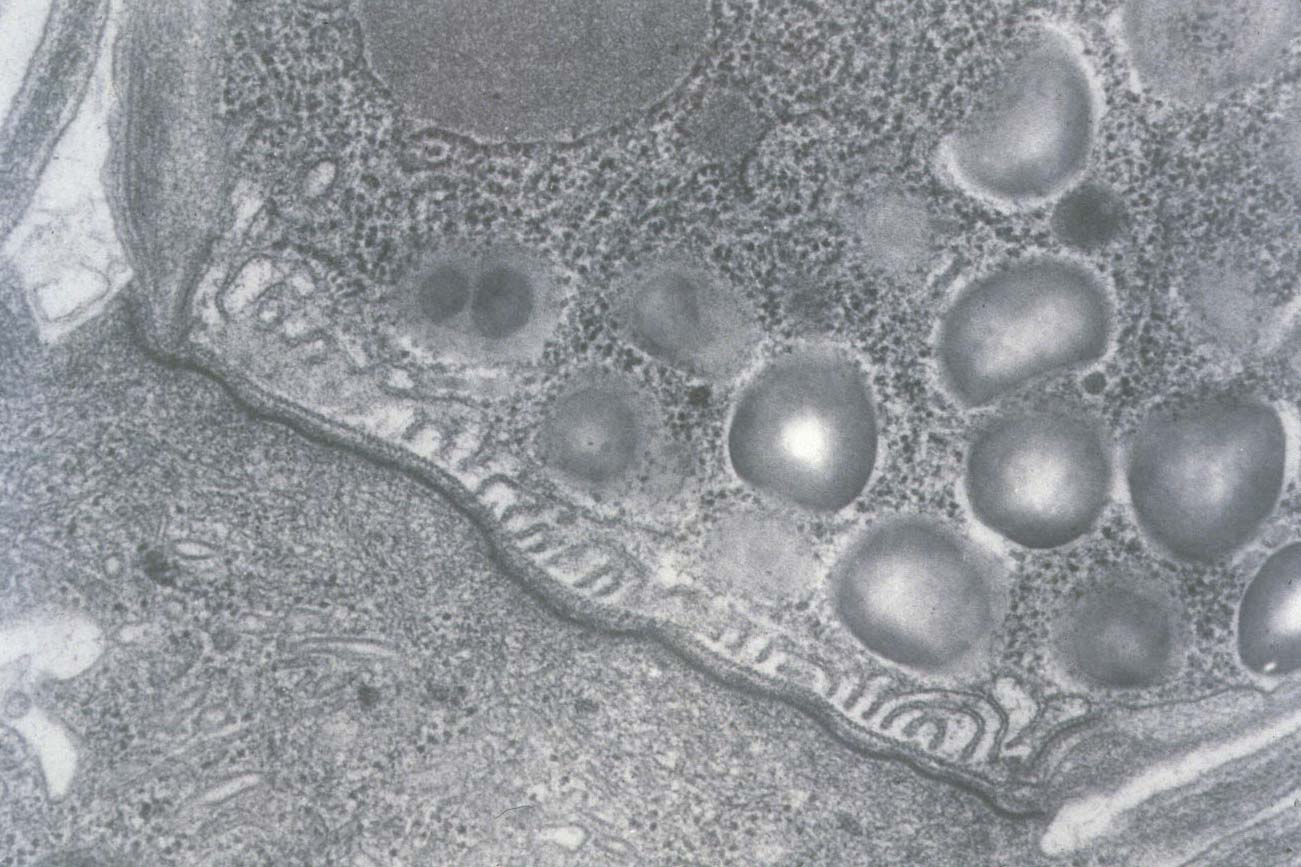
The electron micrograph on the left is of the desmosome-like junction and feeder organelle of Cryptosporidium parvum. Developmental stages occur between the microvilli and are attached to epithelial cells. During penetration of host cells by sporozoites or merozoites, an electron-dense layer forms at the point of attachment and is morphologically reminiscent of a desmosome. A thin layer of host cell cytoplasm envelops the parasite, which may be derived from fused microvilli. As the parasite rounds up and begins to differentiate, finger-like folds of the parasite cytoplasm extend to the level of the electron dense desmosome-like layer, and these folds have been termed the "feeder organelle" (1978, Vet Pathol 15: 417-427). It is often postulated that the feeder organelle and desmosome-like junction somehow facilitate nutrient transport by the parasite. Though likely, experimental data to support this is as yet lacking. |
|---|
Original; transmission electron micrograph by S.J. Upton

Home | Search | What's
New | Help | Comments
Kansas State University | Biology Division