Research
The Membrane Transport Physiology Laboratory aims to understand how specialized proteins, transporters and channels, coordinate their activity, thereby facilitating solute and water movement across cell membranes. These fundamental processes govern practically all critical life functions. Our approach toward attaining this goal is to study the structure, function, and cellular organization of transporter and channel proteins.
Several questions are currently of high interest to us:
1) Functional interactions between channels and transporters: For this work, we chose the Cystic Fibrosis Transmembrane Conductance Regulator (CFTR) and a novel transport protein, the sodium-coupled transporter of short chain fatty acids, SLC5A8. SLC5A8 co-resides with CFTR in the apical membranes of many epithelial cells. is This work is targeted at elucidating how CFTR and SLC5A8 associate (and possibly organize) in signaling complexes via protein-protein interaction domains.
2) Structure-function analysis of anion channels and transporters: these studies combine state-of-the-art in silico approaches utilized by our colleague, Dr. Jeff Comer, with real-time physiological measurements of function established in the Fong Laboratory. For these studies, LRRC8 channels and CLC transporters are starting points for our models.
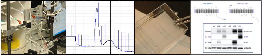
Diverse cell physiological, protein biochemical and molecular biological methods are employed in the Laboratory's research.The Laboratory benefits from state-of-the-art Core facilities offered by the Department of Anatomy and Physiology and the KSU Center "Epithelial Function in Health and Disease".
We are always looking to engage motivated students in our many research projects. Learn about our Lab Members, the Lab's current research focus and Dr. Fong on this site.
Banner: Confocal microscopic image shows anti-zona occludens-1 (ZO-1; green) staining of tight junctions between cultured pig thyroid follicular epithelial cells.
