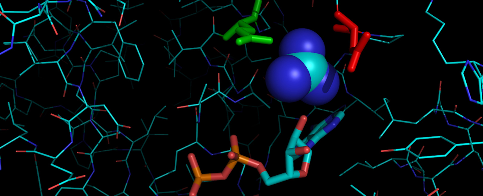
Biochemistry and Molecular Biophysics
Biochemistry: The chemistry of life.
Biochemistry at K-State
Biochemistry provides the foundation for understanding every biological process. To prepare students for careers in medicine, agriculture, biotechnology, and other rapidly evolving industries, our undergraduate and graduate programs are designed to offer exceptional flexibility and versatility.

The department offers Bachelor of Arts and Bachelor of Science degrees in biochemistry or medical biochemistry, as well as a Bachelor of Science in biochemistry and molecular biophysics.
The K-State graduate program in biochemistry and molecular biophysics is an interdisciplinary program including 22 faculty mentors representing six departments. Both a master's and a doctorate level option are available.

Scholarships are awarded annually, and many are renewable.
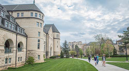
Chalmers Hall
Home of cutting-edge research